Tuesday, April 21, 2015

5. The Deep Front Arm Line

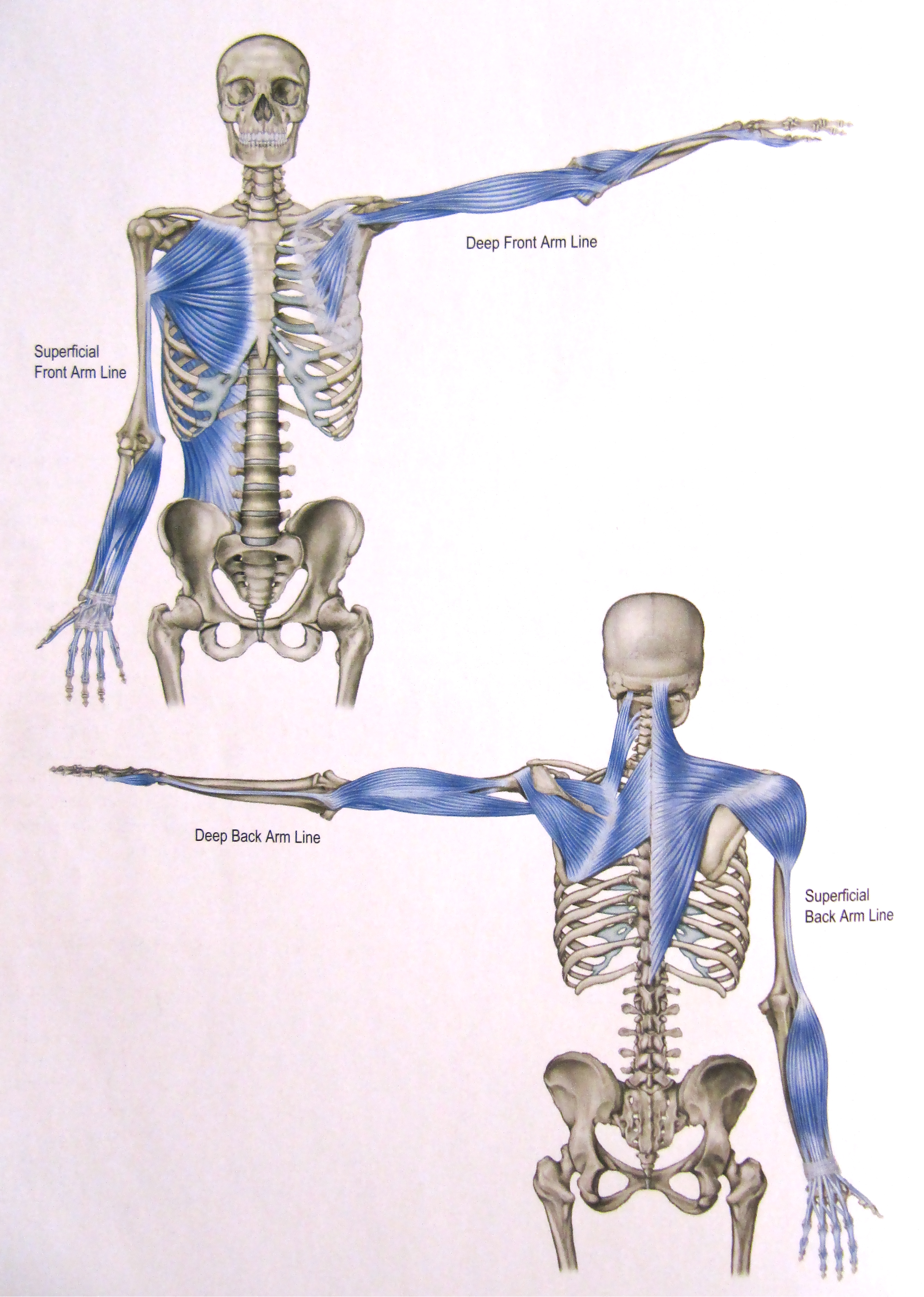
The Deep Front Arm Line


The Arm Lines are, posturally speaking, a bit different from the other myofascial meridians.  The Deep Front Arm Line is a stabilizing line; in poses like the yoga plank, it manages side to side movement of the upper body.  In the open movement of the arm, the DFAL controls the angle of the hand, generally through the thumb, as well as the thumb's grip.
 
- - - - - 


Muscles of the DFAL:
Pectoralis minor
Biceps brachii
Thenar muscles
 
 
Connective tissue (ligaments, tendons, fascial sheets, etc.) of the DFAL:
Clavipectoral fascia
Radial periosteum - anterior border
Radial collateral ligmanets
 
Bony Landmarks of the DFAL:
3rd, 4th, 5th ribs
Coracoid process
Radial tuberosity
Styloid process of radius
Scaphoid, trapezium
Outside of thumb


Common postural deviations within/caused from restriction of the DFAL:
Restriction in the upper rib movement with inhalation
Trouble flexing the arm and lifting from the shoulder to reach upwards
Anterior tilt of the scapula on the ribs - rounded shoulders

Stretches for the DFAL:

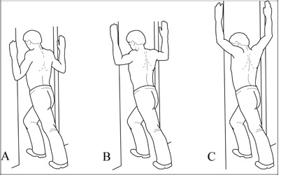
Hands clasped behind your back, shoulders down
 
Doorway stretches for both pectorals major and minor

 
Next week, I'll be covering the Superficial Front Arm Line!
 
- - - - -
 
Take care of yourselves and stretch those arms! :)

Tuesday, April 14, 2015

4. The Spiral Line

The Spiral Line
 
 
The Spiral Line wraps around the body in two helices, right and left, connecting each side of the skull across the upper back top the opposite shoulder, then around to the front of the ribs to cross again at the navel, attaching at the hips.  From there, the SPL passes along the anterolateral (anterior lateral) thigh and across the shin to the arch of the foot, where it then wraps under the foot and runs up along the posterior lateral side of the leg to the ischium and into the erector spine myofascia, ending just about where it started, back at the occipital ridge of the skull.

Postural function of the SPL is to, like most of the myofascial meridians, maintain balance.  The SPL, however, maintains balance across ALL planes.  It mediates rotations in the body and works to steady the truck and leg to keep it from folding into rotational collapse.

Muscles of the SPL:
Splenius capitis and cervicis
Rhomboids major/minor
Serratus anterior
External oblique
Internal oblique
Tensor fasciae latae
Tibialis anterior
Fibularis longs
Bicep memories
Erector spine

(this picture shows half of the spiral line muscles, just so you can see the spiral pattern from one side)

Connective Tissue (ligmanets, tendons, fascial sheets, etc.) of the SPL:
Abdominal aponeurosis
Linea alba
Iliotibial tract
Sacrotuberous ligament
Sacrolumbar fascia

Bony Landmarks that serve as anchors for the SPL:
Occipital ridge
Mastoid process atlas/axis transverse processes
Lower cervical/Upper thoracic spinous processes
Medial border of the Sapulpa
Lateral ribs
Illiac crest/ASIS
Lateral tibia condyle
1st metatarsal base
Fibular head
Ischial tuberosity
Sacrum
Occipital ridge


Common postural deviations: 
Imbalance between left and right
Twisting of the shoulders or hips
Lateral shifts in the body

Stretches for the SPL:

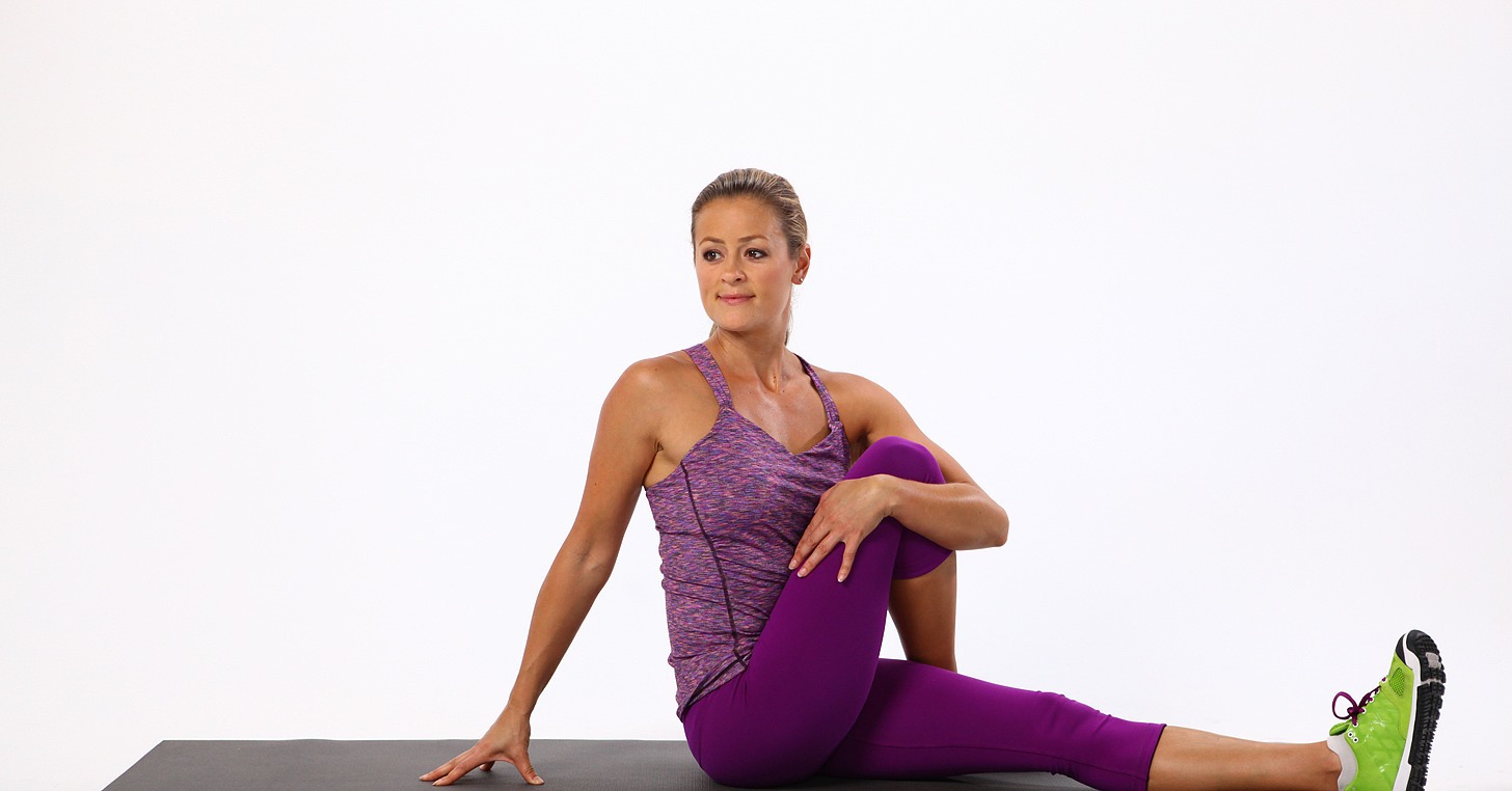
Seated Twist
 
Triangle Pose


- - - - -
 
Next week, I'll start going over the four arm lines, starting with the Deep Front Arm Line!
 
Be good to yourselves! :)

Monday, April 6, 2015

3. The Lateral Line

     Ok! As promised, I'm going over the Lateral Line today.  This particular myofascial meridian assists in (you guessed it!) lateral flexion of the torso, as well as abduction at the hips and eversion of the foot.  It also functions posturally to balance the front and back, and bilaterally to balance left and right.

If you've ever had a health practitioner tell you your hips were off or one hip was high than the other, you can bet this line has some restriction somewhere.

I have several clients (and myself included) who struggle with low back pain due to tightness through the rib, lumbar, and hip segments of this line.
 - - - - -

The Lateral Line


Muscles within the Lateral Line:
Splenius capitis
Sternocleidomastoid
External + interal intercostals
Lateral abdominal obliques
The Abductor muscle group:
  • Gluteus maximus
  • Gluteus medius
  • Gluteus minimus
  • Tensor fasciae latae
The Fibulari muscle group:
  • Peroneus longus
  • Peroneus brevis
  • Peroneus tertius


Connective tissue (tendons, ligaments, fascial sheets) that make up the remainder of the Lateral Line:
Iliotibial tract (IT band)
Anterior ligament of the head of the fibula

Bony landmarks that serve as anchors for the Lateral Line:
Occitpital ridge
Mastoid process
1st + 2nd ribs
Illiac crest
ASIS (Anterior superior iliac spine)
PSIS (Posterior superior iliac spine)
Lateral tibial condyle
Fibular head
1st + 5th metatarsal bases

 
Common postural deviations associated with the Lateral Line:
Ankle pronation/supination
Limited range of motion (particularly in dorsiflexion) of the ankle
Lumbar compression
Shoulder restriction due to over involvement with head stability (I.e. forward head posture)
 
Stretches for the LL:
 
Half moon pose

 
Triangle pose (+modified half moon pose)

 Gate pose
 
So, try these out if you have low back pain, stiffness in the ankles/shoulders, and as always:
 
Be good to yourselves. :)


Tuesday, February 17, 2015

2. The Superficial Front Line

     Last week, I covered the Superficial Back Line, which gives the body its natural primary and secondary curves from top to bottom, so this week I'm going over the SBL's anterior partner:
 The Superficial Front Line

For anyone who suffers from lower back/hip pain, tight/sore ankles, restricted diaphragmatic breathing, or tension headaches from forward head posture, chances are you might have some imbalance throughout your SFL.

- - - - - 

The Superficial Front Line runs along the anterior length of the body in two sections: from the tops of the toes to the anterior, lateral pelvis + from the pubic bone to the head.  This particular track is what gives balance to the Superficial Back Line.  In the picture below, you can see how the posture is affected when the SBL and SFL become unbalanced.

The postural function of the SFL is to allow flexion of the torso and hips, knee extension, and dorsiflexion (pulling upward) of the foot.

Muscles within the SFL:
Sternocleidomastoid
Sternalis
Rectus abdominis
Rectus femoris/quadriceps
Short + long toe extensors
Tibialis anterior

Connective tissue (ligaments, tendons, fascial sheets) that make up the rest of the SFL:
Scalp fascia
Sternochondral fascia
Subpatellar tendon

Bony landmarks the SFL anchors to:
Mastoid process
Sternal manubrium
5th rib
Pubic tubercle
Anterior inferior iliac spine (AIIS)
Patella
Tibial tuberosity
Dorsal surface of toe phalanges


Common postural deviations associated with the SFL:
Limited range of motion in ankle flexion
Anterior pelvic tilt (which can also been seen in the Superficial Back Line, when the erectors of the lumbar spine become shortened)
Forward head posture
Restriction of the diaphragm through the anterior ribs

SFL Stretches:
Cobra pose
Leaning back into full hip extension (the second stretch pictured)
Bridge pose

Camel pose

Backbends are the closest stretch you can do to reach a full stretch throughout the whole SFL.  You can always lay on your back, stretched out over an exercise ball (which I'm about to go do after being at the computer most of the day!)

- - - - -

Next week, I'll cover the Lateral Line.

Be good to yourselves. :)


Wednesday, February 4, 2015

1. The Superficial Back Line

Welcome to the first myofascial meridian!  

The Superficial Back Line

     I personally struggle with this guy.  Tight calves (gastrocnemius), knotted-up hamstrings, shortened erectors in the lumbar area, which means - you guessed it - all the connective tissue (fascia) that encase those muscles and helps keep them tacked down the my bones is nice and tied up, too.  I can definitely thank the Superficial Back Line for my low back issues.

So, if you have pain or tightness along your spine, tightness in the back of your neck, or have trouble touching your toes when you're doing all those awesome stretches I've suggested, you can bet that issues lie within the SBL.

Let's go ahead and go over all the bits and pieces of the SBL so we can get right to the solution to those possible issues.

- - - - -

     The superficial back line (SBL) runs along the entire posterior surface of the body, from the toes all the way up to the brow.  It's postural function is to keep the body upright.  It is what allows us to sit up straight and walk tall.


Each meridian consists of muscles + connective tissues (i.e. ligaments, tendons, fascial sheets) and attach to specific bony landmark to allow mobility through the entire track. 


Muscles within the SBL:
Erector Spinae
Hamstrings
Gastrocnemius
Short toe flexors


Connective tissue (ligaments, tendons, fascial sheets) that make up the rest of the SBL:
Epicranal fascia
Sacrotuberousligament
Achilles tendon
Plantar fascia

The bony landmarks that the SBL anchors to:
Frontal bone
Suboccipital ridge
Occipital ridge
Scarum
Ischial tuberosity
Condyles of the femur
Calcaneus
Plantar surface of the toes


Common postural deviations associated with the SBL are:
Hyperextension of the knees
Lordosis
Hamstring shortness
Hyperextension of the upper cervial spine


Some stretches that address parts of and the entire SBL:
Seated forward bend (A)
Forward bend (B)
Downward dog (C)
Plow pose (D)
Child's pose (not pictured)


Also, laying on your stomach stretched over a large exercise ball (like so) aids in relaxation of the entire SBL.


- - - - -

So...?  How was that?  Do you guys have a better idea of what myofascial meridians are now?
Shoots me any questions or comments you might have here or here (Facebook) or here (Twitter).

Next week, I'll be going over the Superficial Front Line!

Sunday, January 25, 2015

Intro to Myofascial Meridians

I've briefly touched on fascia and myofascial meridians in some of my past blogs, and if you're a client, we've probably talked about them!  So, with the new year, I thought I'd start 2015 off with a 13 part blog series!  You read that right.  
Thirteen. Part. Blog. Series.

Part One: Intro to Myofascial Meridians


What is a myofascial meridian?
     A connected string of myofascial or fascial structures.  Lines that transmit strain and rebound, facilitate movement, and provide stability throughout the structures of our bodies.

How many myofascial meridians exist?
     According to Myers (author of Anatomy Trains), there are twelve specific fascial lines throughout the body.
  1. SBL - superficial back line
  2. SFL - superficial front line
  3. LL - lateral line
  4. SL - spiral line
  5. DFAL - deep front arm line
  6. SFAL - superficial front arm line
  7. DBAL - deep back arm line
  8. SBAL - superficial back arm line
  9. BFL - back functional line
  10. FFL - front functional line
  11. IFL - ipsilateral functional line
  12. DFL - deep front line
Why are myofascial meridians important?
     They make up the "fascial net", which holds everything together.  Connective tissue (fascia) binds every cell to the next and even bonds the internal network of each cell to the mechanical state of the entire body.  These meridians are able to store and communicate information throughout the body as a whole.
   
What are some benefits of addressing postural compensations (postural deviations that cause pain - local or widespread) throughout these myofascial meridians?
  • Skeletal alignment and support
  • Resilience throughout the body
  • Increased range of motion
  • Reduced pain
  • Length throughout the body leading to reduced effort and stress in standing or moving

Each week, I'll be posting an individual article for each myofascial meridians.  I'll go over the muscles, bony landmarks, and common postural compensations associated with each one, as well as stretches and tips to help alleviate related pain.

Stay tuned next week for an in depth look at the Superficial Back Line!

Wednesday, October 29, 2014

Is it Carpal Tunnel Syndrome?

     With all the work we do on our smart phones, tablets, and computers, it's not surprising that many of us suffer from pain in our arms, hands, and fingers.  It's easy to write off the swelling, numbness, tingling, and general discomfort as carpal tunnel syndrome.  However, I've worked with several clients who had surgery for such a diagnosis with little to no relief.  So if it's not CTS, what's the problem?

First, let's clarify what Carpal Tunnel Syndrome is:
     The carpal tunnel is an opening in the wrist made up of the carpal bones, ligaments, and other fibrous tissues.  With Carpal Tunnel Syndrome, the median nerve and tendons traveling through this passageway become compressed when the carpal tunnel is swollen or restricted.  This compression causes numbness and tingling of the wrists, hands, and fingers.

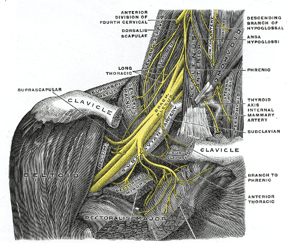
     More commonly though, the impingement of the thoracic outlet (the opening between your collarbone and upper ribs where the nerves and blood vessels pass through to supply the arms and hands) is actually the root of all these symptoms.  When the muscles (i.e. the scalenes) around this opening become tight and shortened, they pull the first rib up against the collarbone, restricting blood, lymph, and nerve supply to the arms, which ultimately causes all the same symptoms as carpal tunnel syndrome.  (The pectoralis minor, triceps, brachialis, and some of the forearm muscles can also become tight and impinge nerves of the arms and hands.)

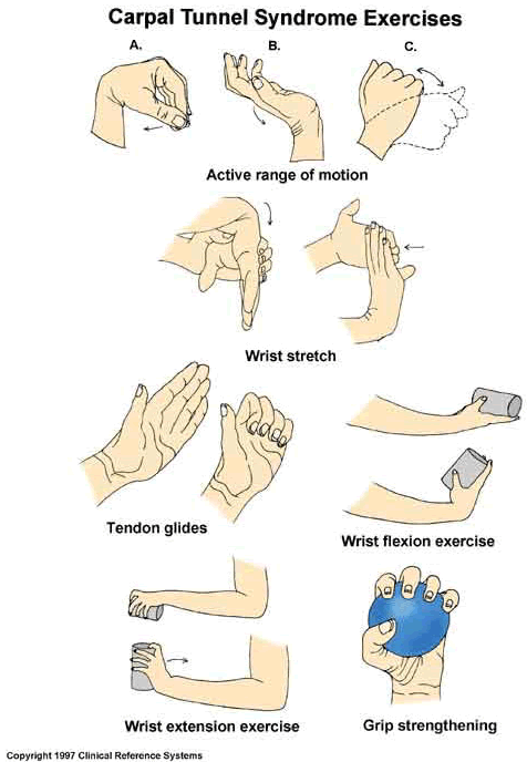
If it is truly CTS, where most of the restriction is at the wrist, maintaining proper body mechanics, stretching your hands and forearms throughout the day, and avoiding poor sleep habits (like bending your wrists or tucking them under your pillow all night) should alleviate many of the symptoms.

I also use contrast bathes frequently for my forearms and hands.  Ice water submersion for 1-2 minutes, then hot water for the same - just go back and forth about 3 to 5 times, always ending with the ice water!  This basically creates a forced circulation, pushing blood out with the ice water then opening up the blood vessels and tissue to new oxygenated blood with the hot.  It works wonders.

If your symptoms seem to be stemming from further up the arm - maybe even into the shoulder and neck - your scalenes (anterior, medial, and posterior) and pectoralis minor are most likely the main culprits. 

Check out my previous post - Shoulder Pain - for stretches and exercises to alleviate tightness and pain in the shoulders and neck.

On a semi-side note, POSTURE is everything!  Check in with your posture throughout the day; you'll probably be amazed by how curved forward and curled up you can get while engrossing yourself in work (or a Netflix binge).  Sit up straight, lift the crown of your head to the ceiling, and let those shoulders fall back.  This might seem easier said than done, but with massage, self care, and stretching, you can absolutely work your way to less pain with better posture.

And I'll leave you with this.  :)