Showing posts with label connective tissue. Show all posts

Tuesday, June 2, 2015

9. The Functional Lines

     Ok, I was originally going to break the Functional Lines into three sections over three weeks, but because of their movements and postural support being so intertwined, I've decided to just cover all three this week.
- - - - -
 
The Functional Lines are meridians generally employed during athletic movements.  They are the least used lines when it comes to standing posture and actually don't have much of an opportunity to shorten or tighten during normal daily activities.  The only postural deviations associated with these lines are a drawing down of one shoulder to the opposite hip in anterior or posterior rotation.
 
- - - - -

 
  The Back Functional Line
 
Muscles:Latissimus dorsi
Gluteus maximus
Vastus lateralis

Connective tissues:Lumbodorsal fascia
Sacral fascia
Subpatellar tendon

Bony landmarks:
Shaft of the humerus
Sacrum
Shaft of the femur
Patella
Tuberosity of the tibia


 The Front Functional Line 
 
Muscles:
Lower edge of pectoralis major
Adductor longus

Connective tissues:Lateral sheath of rectum abdominus

Bony landmarks:Shaft of the humerus
5th, 6th rib cartilage
Pubic tubercle and symphysis
Linea aspera of femur


     The Ipsilateral Functional Line 
Muscles:
Outer edge of the latissimus dorsi
External oblique
Sartorius

Connective tissues:N/A for this line

Bony landmarks:
Shaft of the humerus
End of ribs 10-12
Anterior Superior Iliac Spine
Pes anserinus, medial tibia condyle

Because these lines have little influence on standing posture, there aren't many stretches to suggest, but if you were a tennis player and do have one shoulder drawing closer to the opposite shoulder in rotation, I'd recommend just trying to balance the body out with the opposite rotation ( i.e. if your rotated down and to the right, try to spend some extra time resting in left rotation).

- - - - -
 
So next week will be the last of the Myofascial Meridian series, ending with the Deep Front Line.
 
Be thinking of any topics you would like covered and shoot me an email at laurachancelmt@yahoo.com or comment below on any blog posts!
 
Be good to yourselves. <3


Monday, May 18, 2015

8. The Superficial Back Arm Line

     The Superficial Back Arm Line is the fascial connection from spine to fingers.  It controls arm movements behind our lateral midline (ex. A backhand tennis shot) but, for the most part, limits and contains the work of the Superficial FRONT Arm Line.  The SBAL also maintains control during abduction (lifting away from the body's midline) of the shoulder and arm; this can cause the line to get overworked when the rib cage or spine moves out of alignment with the shoulder girdle.


- - - - -
 
Muscles of the SBAL:
Trapezius
Deltoid
Extensor group
     Brachialis
     Extensor carpi radialus longus/brevis
     Extensor digitorum
     Extensor digiti minimi
     Extensor carpi ulnaris
     Anconeus
 
Connective tissues of the SBAL:
Lateral intermuscular septum
 
Bony landmarks of the SBAL:
Occipital ridge
Nuchal ligament
Thoracic spinous processes
Spine of scapula
Acromion
Lateral third of clavicle
Deltoid tubercle of humerus
Lateral epicondyle of humerus
Dorsal surface of fingers
 
 
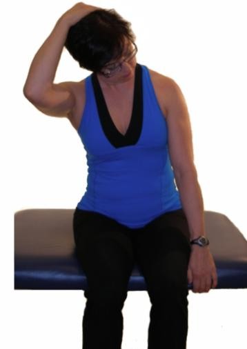
Stretches for the SBAL:
 
 
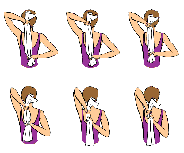
Upper Trapezius Stretch
 
Anterior Deltoid Stretch
 
Medial/Posterior Deltoid Stretch
 
Arm Extension Stretch
 
- - - - -
 
Next week, I'll start The Functional Lines with The Back Functional Line.
 
Stretch, hydrate, breathe deeply, and as always -
Be good to yourselves. <3
 

Monday, May 11, 2015

7. The Deep Back Arm Line


     The Deep Back Arm Line is similar to the Lateral Line in the leg.  It works with the Deep Front Arm Line to adjust the angle of the elbow, as well as limit or allow side-to-side movement of the upper body when in a crawl position, and provide stability from the lateral edge of the hand to the posterior shoulder.


Muscles of the DBAL:
Rhomboids
Rotator cuff muscles:
     Supraspinatus
     Infraspinatus
     Teres minor
     Subscapularis
Triceps brachii
Hypnothenar muscles:
     Abductor digiti minimi
     Flexor digiti minimi brevis
     Opponens digiti minimi


Connective tissues of the DBAL:
Fascia along ulnar periosteum
Ulnar collateral ligaments

Bony landmarks of the DBAL:Spinous process of lower cervical and thoracic vertebrae
C1-4 Transverse processes
Medial border of the scapula
Head of the humerus
Olecranon of the ulna
Triquetrum, hamate
Outside of the little finger


Stretches for the DBAL:

Tricep stretch
 
Ulnar nerve stretch
 
 
- - - - -
 
Stayed tuned in for next week's post - the last of the arm lines: The Superficial Back Arm Line.
 
And I'll leave you all with this:
 
My sweet Bear helping me with my research.
 
 
Take care of yourselves. <3


Monday, May 4, 2015

6. The Superficial Front Arm Line

     The Superficial Front Arm Line controls the positioning of the arm in its lateral and anterior movements.  The larger muscles of the SFAL (the pectoralis major and latissimus dorsi) aid in the force for addiction and extension, movements used in activities like swimming or tennis.  Through the fingers and wrists, the SFAL assists the DFAL in grip.

 
- - - - -

Muscles of the SFAL:
Pectorals major
Latissimus dorsi (not completely pictured below)
Flexor group


Connective tissue of the SFAL:
Medial intermuscular septum
Carpal tunnel

Bony landmarks of the SFAL:
Medial third of clavicle
Coastal cartilage
Lower ribs
Thoracolumbar fascia
Iliac crest
Medial humeral line
Medial humeral epicondyle
Palmar surface of the fingers

 
Common postural deviations associated with the SFAL:
Carpal tunnel impingement
Protracted or rounded shoulders
Finger/hand pain

Stretches for the SFAL:
 

This is a really good representation of how to properly do Downward Facing Dog.
 
- - - - -
 
Sorry I missed a week, guys; Austin's allergies had it in for me, but thanks to lots of sleep and NetiPot, I'm back online.  I hope you are well!
 
I'll be back next week with the Deep Back Arm Line.
 
Be good to yourselves. <3

Tuesday, April 14, 2015

4. The Spiral Line

The Spiral Line
 
 
The Spiral Line wraps around the body in two helices, right and left, connecting each side of the skull across the upper back top the opposite shoulder, then around to the front of the ribs to cross again at the navel, attaching at the hips.  From there, the SPL passes along the anterolateral (anterior lateral) thigh and across the shin to the arch of the foot, where it then wraps under the foot and runs up along the posterior lateral side of the leg to the ischium and into the erector spine myofascia, ending just about where it started, back at the occipital ridge of the skull.

Postural function of the SPL is to, like most of the myofascial meridians, maintain balance.  The SPL, however, maintains balance across ALL planes.  It mediates rotations in the body and works to steady the truck and leg to keep it from folding into rotational collapse.

Muscles of the SPL:
Splenius capitis and cervicis
Rhomboids major/minor
Serratus anterior
External oblique
Internal oblique
Tensor fasciae latae
Tibialis anterior
Fibularis longs
Bicep memories
Erector spine

(this picture shows half of the spiral line muscles, just so you can see the spiral pattern from one side)

Connective Tissue (ligmanets, tendons, fascial sheets, etc.) of the SPL:
Abdominal aponeurosis
Linea alba
Iliotibial tract
Sacrotuberous ligament
Sacrolumbar fascia

Bony Landmarks that serve as anchors for the SPL:
Occipital ridge
Mastoid process atlas/axis transverse processes
Lower cervical/Upper thoracic spinous processes
Medial border of the Sapulpa
Lateral ribs
Illiac crest/ASIS
Lateral tibia condyle
1st metatarsal base
Fibular head
Ischial tuberosity
Sacrum
Occipital ridge


Common postural deviations: 
Imbalance between left and right
Twisting of the shoulders or hips
Lateral shifts in the body

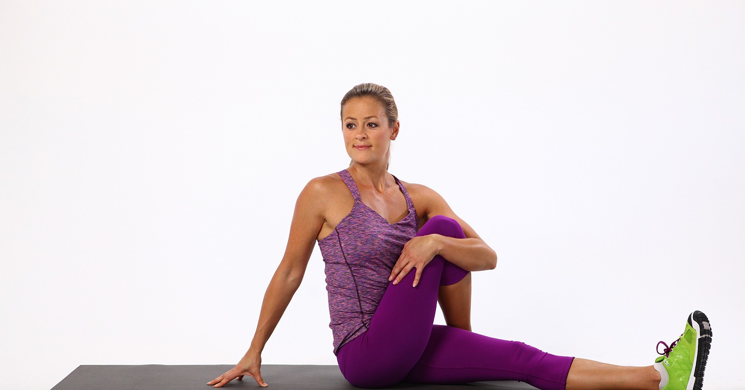
Stretches for the SPL:

Seated Twist
 
Triangle Pose


- - - - -
 
Next week, I'll start going over the four arm lines, starting with the Deep Front Arm Line!
 
Be good to yourselves! :)

Monday, April 6, 2015

3. The Lateral Line

     Ok! As promised, I'm going over the Lateral Line today.  This particular myofascial meridian assists in (you guessed it!) lateral flexion of the torso, as well as abduction at the hips and eversion of the foot.  It also functions posturally to balance the front and back, and bilaterally to balance left and right.

If you've ever had a health practitioner tell you your hips were off or one hip was high than the other, you can bet this line has some restriction somewhere.

I have several clients (and myself included) who struggle with low back pain due to tightness through the rib, lumbar, and hip segments of this line.
 - - - - -

The Lateral Line


Muscles within the Lateral Line:
Splenius capitis
Sternocleidomastoid
External + interal intercostals
Lateral abdominal obliques
The Abductor muscle group:
  • Gluteus maximus
  • Gluteus medius
  • Gluteus minimus
  • Tensor fasciae latae
The Fibulari muscle group:
  • Peroneus longus
  • Peroneus brevis
  • Peroneus tertius


Connective tissue (tendons, ligaments, fascial sheets) that make up the remainder of the Lateral Line:
Iliotibial tract (IT band)
Anterior ligament of the head of the fibula

Bony landmarks that serve as anchors for the Lateral Line:
Occitpital ridge
Mastoid process
1st + 2nd ribs
Illiac crest
ASIS (Anterior superior iliac spine)
PSIS (Posterior superior iliac spine)
Lateral tibial condyle
Fibular head
1st + 5th metatarsal bases

 
Common postural deviations associated with the Lateral Line:
Ankle pronation/supination
Limited range of motion (particularly in dorsiflexion) of the ankle
Lumbar compression
Shoulder restriction due to over involvement with head stability (I.e. forward head posture)
 
Stretches for the LL:
 
Half moon pose

 
Triangle pose (+modified half moon pose)

 Gate pose
 
So, try these out if you have low back pain, stiffness in the ankles/shoulders, and as always:
 
Be good to yourselves. :)


Wednesday, February 4, 2015

1. The Superficial Back Line

Welcome to the first myofascial meridian!  

The Superficial Back Line

     I personally struggle with this guy.  Tight calves (gastrocnemius), knotted-up hamstrings, shortened erectors in the lumbar area, which means - you guessed it - all the connective tissue (fascia) that encase those muscles and helps keep them tacked down the my bones is nice and tied up, too.  I can definitely thank the Superficial Back Line for my low back issues.

So, if you have pain or tightness along your spine, tightness in the back of your neck, or have trouble touching your toes when you're doing all those awesome stretches I've suggested, you can bet that issues lie within the SBL.

Let's go ahead and go over all the bits and pieces of the SBL so we can get right to the solution to those possible issues.

- - - - -

     The superficial back line (SBL) runs along the entire posterior surface of the body, from the toes all the way up to the brow.  It's postural function is to keep the body upright.  It is what allows us to sit up straight and walk tall.


Each meridian consists of muscles + connective tissues (i.e. ligaments, tendons, fascial sheets) and attach to specific bony landmark to allow mobility through the entire track. 


Muscles within the SBL:
Erector Spinae
Hamstrings
Gastrocnemius
Short toe flexors


Connective tissue (ligaments, tendons, fascial sheets) that make up the rest of the SBL:
Epicranal fascia
Sacrotuberousligament
Achilles tendon
Plantar fascia

The bony landmarks that the SBL anchors to:
Frontal bone
Suboccipital ridge
Occipital ridge
Scarum
Ischial tuberosity
Condyles of the femur
Calcaneus
Plantar surface of the toes


Common postural deviations associated with the SBL are:
Hyperextension of the knees
Lordosis
Hamstring shortness
Hyperextension of the upper cervial spine


Some stretches that address parts of and the entire SBL:
Seated forward bend (A)
Forward bend (B)
Downward dog (C)
Plow pose (D)
Child's pose (not pictured)


Also, laying on your stomach stretched over a large exercise ball (like so) aids in relaxation of the entire SBL.


- - - - -

So...?  How was that?  Do you guys have a better idea of what myofascial meridians are now?
Shoots me any questions or comments you might have here or here (Facebook) or here (Twitter).

Next week, I'll be going over the Superficial Front Line!

Monday, October 20, 2014

IT Band Syndrome

     I got this awesome (and incredibly relevant) topic choice from one of my FB followers!  With this amazing cool(er) weather, I've starting to run again, which means  - you guessed it! -  lots of foam rolling and stretching!  Unfortunately, that doesn't always cut it.  I have several of cyclist and runner clients who have chronically tight legs and hips, but more specifically, TIGHT I.T. BANDS!

The Iliotibial Tract (IT Band) is a fibrous strip of thick connective tissue (a reinforcement of the fascia latte: the deep connective tissue of the thigh).  It originates at the iliac crest (the curve of your hip bone) and runs aloing the lateral side of the thigh, insterting at the top of the tibia (the shin).  It encases the Tensor Fascia Latte (TFL) and also recieves most of the gluteus maximus tendon.

Actions: stabilizes the knee in extension and in partial flexion (i.e. walking/running, squatting, cycling, etc.)

Iliotibial band syndrome (ITBS or ITBFS, for iliotibial band friction syndrome, Runner's Knee) is the chronic inflammation of the IT Band; it is one of the most common causes of lateral knee pain.

There are several different things that can cause ITBS, such as overuse, leg length discrepancies, and improper form when exercising/running. 

So what can we do for ITBS?
As far as massage goes, trigger point and myofascial techniques would be the way to go to alleviate some tightness throughout the entire thigh, with a focus on the knee attachment points of the IT Band and hamstring tendons (just behind the IT Band insertion point).  The TFL and the Gluteus Maximus would be the next section to address because of their direct relation to the IT Band.

Your Homework:
For prevention and management, stretching and proper strengthening exercises for the lateral thigh will take strain off the IT Band.


For more exercises/stretches for the legs, follow me on Pinterest.

And if you have a flair up, always R.I.C.E. (Rest, Ice, Compress, Elevate).  
Really.  Do NOT power through it.  


Take care of yourself.  :)


Saturday, July 26, 2014

Neck Pain

Hey guys!
I hope you are all doing well, and have had an amazing summer!

     With all the rain happening off and on, everyone’s allergies have been miserable, including mine.  I’d had a horrible headache for almost a week, with NetiPot, Epsom salt baths, and my usual allergy arsenal barely touching it.

     So! I poured over all my reference books for a good while before I turned to YouTube to look for cervical spine stretches (I know I talked about TMJ and Forward Head Posture in one of my previous posts, but I couldn’t even get comfortable enough to do those stretches!)  I finally found this video by Dr. Paula Moore:

     It has worked WONDERS for not only my neck pain, but even pain I was unaware of in my mid back and arms!  After three days of doing her exercise/stretch every time I thought about it or found myself leaning my chin on my hand at the computer, I was loose enough to do these amazing neck stretches.


Do them.  Every time you find yourself craning your neck forward or propping it way too high when watching tv; straighten up, stretch the crown of your head as if you are trying to touch the ceiling, and do.these.stretches.

They will change your cervical spine’s life. :)

You can follow my Relief: Shoulder, Neck, and Head Aches board on Pinterest for more upper back pain solutions!