I recently started doing Pilates. Getting in better shape has been a personal goal lately, especially with the wedding coming up! My general idea was I could tone up and get my posture straightened out, so I go in, talk to my personal pilates instructor, get all the medical history out of the way, and we're off. Or so I thought.
She shows me some of the machines along with the rest of the studio before she leads me over the a wooden rack on the wall with two half balls placed in front of it. She tells me to place my heels on the balls and evenly distribute my weight between my heel and the ball of my foot. Ok. Easy enough.
But once I get my heels up there, I realize how tight the entire sole of each foot was. The instructor patiently walked me through shifting my weight back and forth, side to side, front to back, and we transitioned from heel to arch to ball to toes, all the while, I'm TRYING to keep my weight evenly distributed between my feet.
After a bit, she tells me to try them out. See what it feels like to move; grounded through your feet. Even after having 10 sessions of Rolfing, I had never felt so connected to my feet. It was amazing. I could feel how the tightness in my feet had been affecting my calves up into my knees and hamstrings, even into my hips and low back!
Now, opening my feet is one of the first things I do in the morning and the last thing I do at night - if I'm really good about it, I'll do it in between sessions and throughout the day.
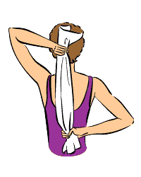
You can use any of these things to open your feet:
Or you can improvise and find whatever works best for you.
Our feet are our connection to the Earth - they are where our support starts against gravity.
The Deep Front Line is the body's myofascial core, as it defines the deep three-dimensional space deep within the body. It's main postural function is providing support and lift in the inner arch of the foot, stabilizing both segments of each leg, as well as the hips, supporting the lumbar spine from the front, giving shape to the abdominopelvic balloon, stabilizing the chest while allowing the expansion and relaxation of breathing, and balancing the neck and head.
If this line is lacking definition, strength, and/or proper balance, there is an overall shortening throughout the body, which encourages collapse of the pelvic and spinal core, laying the groundwork for postural deviations in all the other meridians.
The Deep Front Line can be broken down into six parts:
Lowest Common
Lower Posterior
Lower Anterior
Upper Posterior
Upper Middle
Upper Anterior
So, I've broken down each list below accordingly.
Muscles of the DFL:
Lowest Common:
Tibialis posterior
Long toe flexor
Lower Posterior:
Adductor magnus and minimus Lower Anterior:
Adductor brevis and longus
Psoas
Iliacus
Pectineus
Femoral triangle Upper Posterior:
Longus colli and cavities Upper Middle:
Posterior diaphragm
Pericardium
Mediastinum
Scalene muscles
Upper Anterior:
Anterior diaphragm
Transversus thoracis
Infrahyoid muscles
Suprahyoid muscles
Connective tissues of the DFL:
Lowest Common:
Fasciae of popliteus
Knee capsule Lower Posterior:
Posterior intermuscular septum
Pelvis floor fascia
Levator ani
Obturator internus fascia
Anterior sacral fasciae
Anterior longitudinal ligament Lower Anterior:
Medial intermuscular septum Upper Posterior:
Anterior longitudal ligament Upper Middle:
Crura of the diaphragm
Central tendon
Parietal pleura
Fascia prevertebralis
Pharyngeal raphe
Medial scalene fascia Upper Anterior:
Fascia endothoracica
Fascia pretrachialis
Bony landmarks of the DFL: Lowest Common:
Plantar tarsal bones
Plantar surface of toes
Superior/posterior tibia/fibula
Medial femoral epicondyle Lower Posterior:
Medial femoral epicondyle
Ischial ramus
Coccyx
Lumbar vertebral bodies Lower Anterior:
Medial femoral epicondyle
Linea aspera of femur
Lesser trochanter of femur
Lumbar vertebral bodies and transverse processes Upper Posterior:
Lumbar vertebral bodies
Basilar portion of occiput Upper Middle:
Lumbar vertebral bodies
Basilar portion of occiput
Cervical transverse processes Upper Anterior:
Lumbar vertebral bodies
Posterior surface of subtotal
Cartilage
Xiphoid process
Posterior manubrium
Hyoid bone
Mandible
- - - - -
Pilates is a great way to engage this line properly, therefore setting the foundation for all the other lines to function at their best.
Here's a great video that shows a lunge stretch for the DFL:
- - - - -
And that's it!
We've covered the entire series of myofascial meridian from Thomas Myers' Anatomy Trains - which you can purchase here for a more in depth understanding of these lines!
I'll be back the week of the 22nd with a new topic and great tips on pain relief and healthy living.
Ok, I was originally going to break the Functional Lines into three sections over three weeks, but because of their movements and postural support being so intertwined, I've decided to just cover all three this week.
- - - - -
The Functional Lines are meridians generally employed during athletic movements. They are the least used lines when it comes to standing posture and actually don't have much of an opportunity to shorten or tighten during normal daily activities. The only postural deviations associated with these lines are a drawing down of one shoulder to the opposite hip in anterior or posterior rotation.
- - - - -
The Back Functional Line
Muscles:Latissimus dorsi
Gluteus maximus
Vastus lateralis
Connective tissues:Lumbodorsal fascia
Sacral fascia
Subpatellar tendon
Bony landmarks:
Shaft of the humerus
Sacrum
Shaft of the femur
Patella
Tuberosity of the tibia
The Front Functional Line
Muscles:
Lower edge of pectoralis major
Adductor longus
Connective tissues:Lateral sheath of rectum abdominus
Bony landmarks:Shaft of the humerus
5th, 6th rib cartilage
Pubic tubercle and symphysis
Linea aspera of femur
The Ipsilateral Functional Line
Muscles:
Outer edge of the latissimus dorsi
External oblique
Sartorius
Connective tissues:N/A for this line
Bony landmarks:Shaft of the humerus
End of ribs 10-12
Anterior Superior Iliac Spine
Pes anserinus, medial tibia condyle
Because these lines have little influence on standing posture, there aren't many stretches to suggest, but if you were a tennis player and do have one shoulder drawing closer to the opposite shoulder in rotation, I'd recommend just trying to balance the body out with the opposite rotation ( i.e. if your rotated down and to the right, try to spend some extra time resting in left rotation).
- - - - -
So next week will be the last of the Myofascial Meridian series, ending with the Deep Front Line.
Be thinking of any topics you would like covered and shoot me an email at laurachancelmt@yahoo.com or comment below on any blog posts!
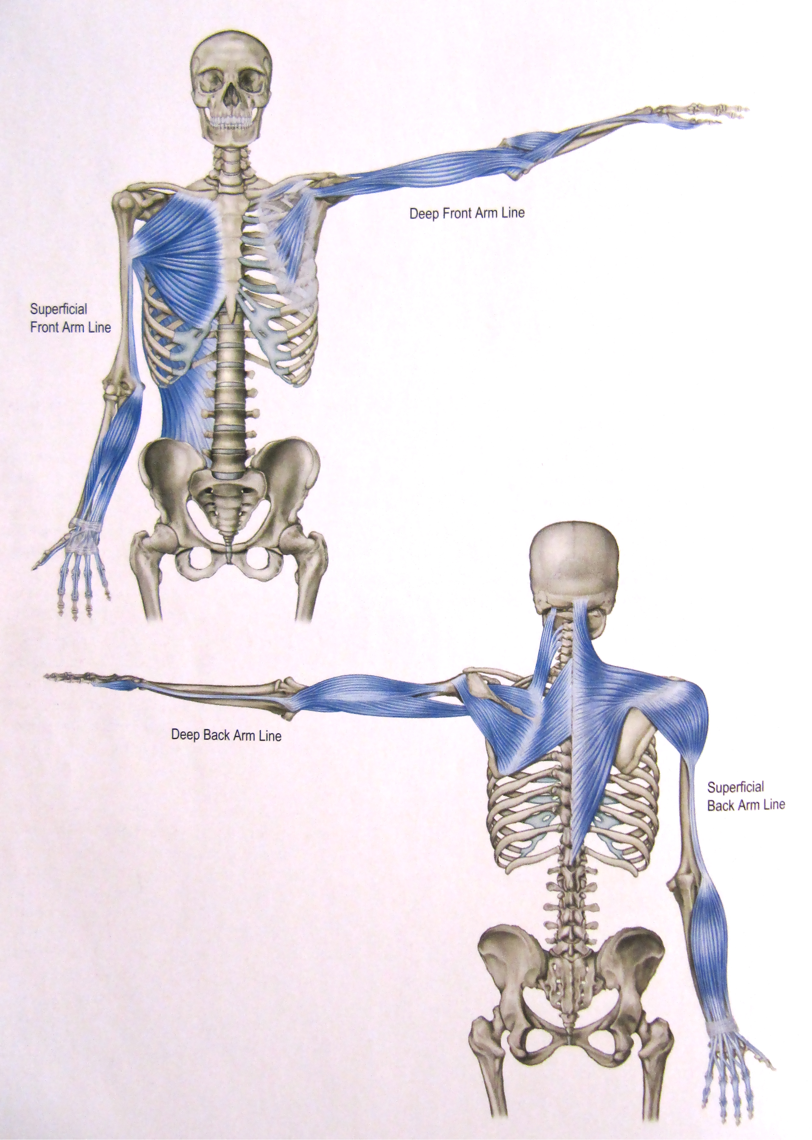
The Superficial Back Arm Line is the fascial connection from spine to fingers. It controls arm movements behind our lateral midline (ex. A backhand tennis shot) but, for the most part, limits and contains the work of the Superficial FRONT Arm Line. The SBAL also maintains control during abduction (lifting away from the body's midline) of the shoulder and arm; this can cause the line to get overworked when the rib cage or spine moves out of alignment with the shoulder girdle.
- - - - -
Muscles of the SBAL:
Trapezius Deltoid
Extensor group
Brachialis
Extensor carpi radialus longus/brevis
Extensor digitorum
Extensor digiti minimi Extensor carpi ulnaris
Anconeus
Connective tissues of the SBAL:
Lateral intermuscular septum
Bony landmarks of the SBAL:
Occipital ridge
Nuchal ligament
Thoracic spinous processes
Spine of scapula Acromion
Lateral third of clavicle
Deltoid tubercle of humerus
Lateral epicondyle of humerus
Dorsal surface of fingers
Stretches for the SBAL:
Upper Trapezius Stretch
Anterior Deltoid Stretch
Medial/Posterior Deltoid Stretch
Arm Extension Stretch
- - - - -
Next week, I'll start The Functional Lines with The Back Functional Line.
The Deep Back Arm Line is similar to the Lateral Line in the leg. It works with the Deep Front Arm Line to adjust the angle of the elbow, as well as limit or allow side-to-side movement of the upper body when in a crawl position, and provide stability from the lateral edge of the hand to the posterior shoulder.
Muscles of the DBAL:
Rhomboids
Rotator cuff muscles:
Supraspinatus
Infraspinatus
Teres minor
Subscapularis
Triceps brachii
Hypnothenar muscles:
Abductor digiti minimi
Flexor digiti minimi brevis
Opponens digiti minimi
Connective tissues of the DBAL:
Fascia along ulnar periosteum
Ulnar collateral ligaments
Bony landmarks of the DBAL:Spinous process of lower cervical and thoracic vertebrae
C1-4 Transverse processes
Medial border of the scapula
Head of the humerus
Olecranon of the ulna
Triquetrum, hamate
Outside of the little finger
Stretches for the DBAL:
Tricep stretch
Ulnar nerve stretch
- - - - -
Stayed tuned in for next week's post - the last of the arm lines: The Superficial Back Arm Line.
The Superficial Front Arm Line controls the positioning of the arm in its lateral and anterior movements. The larger muscles of the SFAL (the pectoralis major and latissimus dorsi) aid in the force for addiction and extension, movements used in activities like swimming or tennis. Through the fingers and wrists, the SFAL assists the DFAL in grip.
- - - - -
Muscles of the SFAL:
Pectorals major
Latissimus dorsi (not completely pictured below)
Flexor group
Connective tissue of the SFAL:
Medial intermuscular septum
Carpal tunnel
Bony landmarks of the SFAL:
Medial third of clavicle
Coastal cartilage
Lower ribs
Thoracolumbar fascia
Iliac crest
Medial humeral line
Medial humeral epicondyle
Palmar surface of the fingers
Common postural deviations associated with the SFAL:
Carpal tunnel impingement
Protracted or rounded shoulders
Finger/hand pain
Stretches for the SFAL:
This is a really good representation of how to properly do Downward Facing Dog.
- - - - -
Sorry I missed a week, guys; Austin's allergies had it in for me, but thanks to lots of sleep and NetiPot, I'm back online. I hope you are well!
I'll be back next week with the Deep Back Arm Line.
The Arm Lines are, posturally speaking, a bit different from the other myofascial meridians. The Deep Front Arm Line is a stabilizing line; in poses like the yoga plank, it manages side to side movement of the upper body. In the open movement of the arm, the DFAL controls the angle of the hand, generally through the thumb, as well as the thumb's grip.
- - - - -
Muscles of the DFAL:
Pectoralis minor
Biceps brachii
Thenar muscles
Connective tissue (ligaments, tendons, fascial sheets, etc.) of the DFAL:
Clavipectoral fascia
Radial periosteum - anterior border
Radial collateral ligmanets
Bony Landmarks of the DFAL:
3rd, 4th, 5th ribs
Coracoid process
Radial tuberosity
Styloid process of radius
Scaphoid, trapezium
Outside of thumb
Common postural deviations within/caused from restriction of the DFAL:
Restriction in the upper rib movement with inhalation
Trouble flexing the arm and lifting from the shoulder to reach upwards
Anterior tilt of the scapula on the ribs - rounded shoulders
Stretches for the DFAL:
Hands clasped behind your back, shoulders down
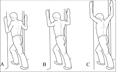
Doorway stretches for both pectorals major and minor
Next week, I'll be covering the Superficial Front Arm Line!
- - - - -
Take care of yourselves and stretch those arms! :)
The Spiral Line wraps around the body in two helices, right and left, connecting each side of the skull across the upper back top the opposite shoulder, then around to the front of the ribs to cross again at the navel, attaching at the hips. From there, the SPL passes along the anterolateral (anterior lateral) thigh and across the shin to the arch of the foot, where it then wraps under the foot and runs up along the posterior lateral side of the leg to the ischium and into the erector spine myofascia, ending just about where it started, back at the occipital ridge of the skull.
Postural function of the SPL is to, like most of the myofascial meridians, maintain balance. The SPL, however, maintains balance across ALL planes. It mediates rotations in the body and works to steady the truck and leg to keep it from folding into rotational collapse.
Muscles of the SPL:
Splenius capitis and cervicis
Rhomboids major/minor
Serratus anterior
(this picture shows half of the spiral line muscles, just so you can see the spiral pattern from one side)
Connective Tissue (ligmanets, tendons, fascial sheets, etc.) of the SPL:
Abdominal aponeurosis
Linea alba
Iliotibial tract
Sacrotuberous ligament
Sacrolumbar fascia
Bony Landmarks that serve as anchors for the SPL:
Occipital ridge
Mastoid process atlas/axis transverse processes
Lower cervical/Upper thoracic spinous processes
Medial border of the Sapulpa
Lateral ribs
Illiac crest/ASIS
Lateral tibia condyle
1st metatarsal base
Fibular head
Ischial tuberosity
Sacrum
Occipital ridge
Common postural deviations:
Imbalance between left and right
Twisting of the shoulders or hips
Lateral shifts in the body
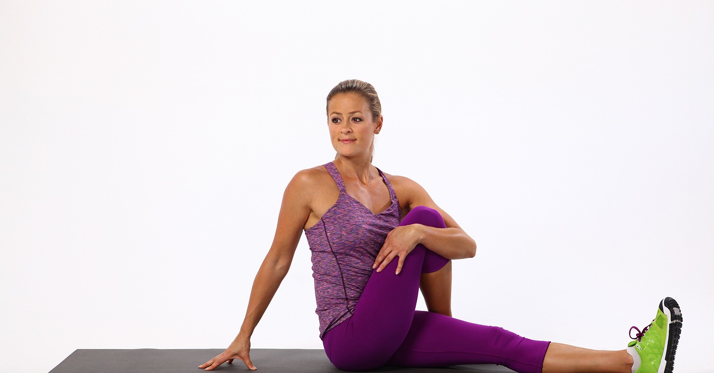
Stretches for the SPL:
Seated Twist
Triangle Pose
- - - - -
Next week, I'll start going over the four arm lines, starting with the Deep Front Arm Line!
Ok! As promised, I'm going over the Lateral Line today. This particular myofascial meridian assists in (you guessed it!) lateral flexion of the torso, as well as abduction at the hips and eversion of the foot. It also functions posturally to balance the front and back, and bilaterally to balance left and right.
If you've ever had a health practitioner tell you your hips were off or one hip was high than the other, you can bet this line has some restriction somewhere.
I have several clients (and myself included) who struggle with low back pain due to tightness through the rib, lumbar, and hip segments of this line.
- - - - -
The Lateral Line
Muscles within the Lateral Line:
Splenius capitis
Sternocleidomastoid
External + interal intercostals
Lateral abdominal obliques
The Abductor muscle group:
Gluteus maximus
Gluteus medius
Gluteus minimus
Tensor fasciae latae
The Fibulari muscle group:
Peroneus longus
Peroneus brevis
Peroneus tertius
Connective tissue (tendons, ligaments, fascial sheets) that make up the remainder of the Lateral Line:
Iliotibial tract (IT band)
Anterior ligament of the head of the fibula
Bony landmarks that serve as anchors for the Lateral Line:
Occitpital ridge
Mastoid process
1st + 2nd ribs
Illiac crest
ASIS (Anterior superior iliac spine)
PSIS (Posterior superior iliac spine)
Lateral tibial condyle
Fibular head
1st + 5th metatarsal bases
Common postural deviations associated with the Lateral Line:
Ankle pronation/supination
Limited range of motion (particularly in dorsiflexion) of the ankle
Lumbar compression
Shoulder restriction due to over involvement with head stability (I.e. forward head posture)
Stretches for the LL:
Half moon pose
Triangle pose (+modified half moon pose)
Gate pose
So, try these out if you have low back pain, stiffness in the ankles/shoulders, and as always:
Last week, I covered the Superficial Back Line, which gives the body its natural primary and secondary curves from top to bottom, so this week I'm going over the SBL's anterior partner:
The Superficial Front Line
For anyone who suffers from lower back/hip pain, tight/sore ankles, restricted diaphragmatic breathing, or tension headaches from forward head posture, chances are you might have some imbalance throughout your SFL.
- - - - -
The Superficial Front Line runs along the anterior length of the body in two sections: from the tops of the toes to the anterior, lateral pelvis + from the pubic bone to the head. This particular track is what gives balance to the Superficial Back Line. In the picture below, you can see how the posture is affected when the SBL and SFL become unbalanced.
The postural function of the SFL is to allow flexion of the torso and hips, knee extension, and dorsiflexion (pulling upward) of the foot.
Muscles within the SFL:
Sternocleidomastoid
Sternalis
Rectus abdominis
Rectus femoris/quadriceps
Short + long toe extensors
Tibialis anterior
Connective tissue (ligaments, tendons, fascial sheets) that make up the rest of the SFL:
Scalp fascia
Sternochondral fascia
Subpatellar tendon
Bony landmarks the SFL anchors to:
Mastoid process
Sternal manubrium
5th rib
Pubic tubercle
Anterior inferior iliac spine (AIIS)
Patella
Tibial tuberosity
Dorsal surface of toe phalanges
Common postural deviations associated with the SFL:
Limited range of motion in ankle flexion
Anterior pelvic tilt (which can also been seen in the Superficial Back Line, when the erectors of the lumbar spine become shortened)
Forward head posture
Restriction of the diaphragm through the anterior ribs
SFL Stretches:
Cobra pose
Leaning back into full hip extension (the second stretch pictured)
Bridge pose
Camel pose
Backbends are the closest stretch you can do to reach a full stretch throughout the whole SFL. You can always lay on your back, stretched out over an exercise ball (which I'm about to go do after being at the computer most of the day!)